Knee Joint Anatomy
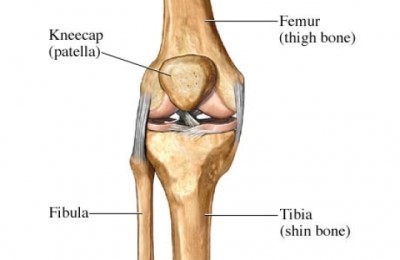
The knee is made up of two joints, the tibiofemoral joint and the patellofemoral joint. The tibiofemoral joint is made up of the femur (thigh bone) and the tibia (shin bone). The patellofemoral joint is made up of the patella (knee cap) and the femur (thigh bone).
Cartilage
Articular cartilage is the tissue that covers the ends of your bones in each joint. There are also two menisci, which are made up of cartilage, in between the femur and the tibia. In the knee, cartilage:
- Helps the bones glide smoothly over each other when you bend or straighten your leg.
- Protects your knee by preventing the bones from rubbing against each other. Cartilage is tough and flexible. But it wears away as you age.
Impact of Arthritis on The Joint
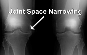
Osteoarthritis is wear and tear of the cartilage layer on the ends of the bones. The space between the two bones see on x-ray is not space, instead, it is cartilage that cannot be seen by the x-ray. As this cartilage wears down the space between the two joints narrow. This can increase pain, swelling, and stiffness.